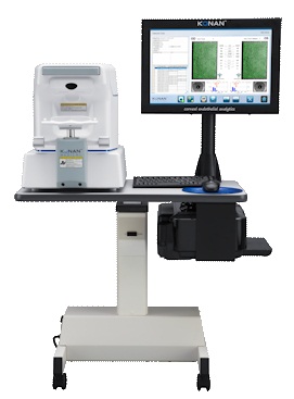
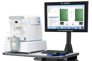
Microscopio Especular CellChek XL
PRESBYOND Laser Blended Vision
Customized. All distances. Immediate.
For more Info, please scroll down.
konan
Highlights
CellChek Konan specular microscopes are the global leaders for specular endothelial analysis, both for routine clinical practice and clinical research.
Simple to use
The CellChek XL specular microscope represents the latest in endothelial cell analysis, algorithms, and computer technology. Enhanced with auto-focus, auto-alignment, and auto-cell counting, the CellChek XL easily captures consistent, high quality images of the patient’s corneal endothelium using a patented method that identifies the position of the cellular interface.
Powerful multi-point, analytic detail
The CellChek XL offers five fixation points for image capture at the center and four peripheral sites, allowing a more comprehensive look at the cornea. This is particularly valuable in cases such as keratoconus | corneal transplantations, DSAEK, CXL, or the presence of corneal dystrophies.
Location specific data samples
Unique to the CellChek XL, the CellChek™ software automatically records the location from which the data samples were acquired. One strong value of specular microscopy is to be able to assess and quantify change in the cornea over time. Without location data,trending is simply not accurate or reliable.
Advanced database features
Fully integrated database management system allows robust data mining and simplification of links to electronic medical records.
Non-contact pachymetry
Using non-contact optical pachymetry, the CellChek XL provides corneal thickness measurements at all five data sample sites. Independent studies have shown this to be more accurate than traditional ultrasound pachymetry.1
The only FDA 510(k) cleared specular microscope («NQE»)
Konan is the only company to have products FDA 510(k) cleared under «Specular Microscopes» Product Code NQE. Why accept anything less than the assurance of the FDA’s review for both imaging and assessment of the corneal endothelial cell layer, morphology of endothelial cells, and corneal pachymetry. Exclusively from Konan.
Flexibility of analysis methods for maximum usability
– Fully automated: minimized examination time of about 15 seconds
– Semi automated: operator can make subjective enhancements to auto-generated cellular overlay
– Center method: Konan’s patented strategy is often used, due to high accuracy, by professional reading centers
– Flex-center method: high value for analyzing difficult images that contain a small number of cells in the sample
Product Description:
The CellChek XL includes specular microscope, analysis software with fully integrated database and trends analysis, and standard accessories: 19″ touchscreen monitor, electric table, and printer.
The Konan CellChek XL provides the optimum side-by-side configuration for minimized use of
valuable clinic floor space and easy patient managment / positioning by the technician.
Konan products are evaluated, certified, and CE marked by TÜV Rhineland Product Safety GmbH of Germany.
Related Download Material
CRS-Master from ZEISS
Planing customized treatments
MEL 90 from ZEISS
Advancing excimer laser technology
MEL 80
High Performance Meets Versatility